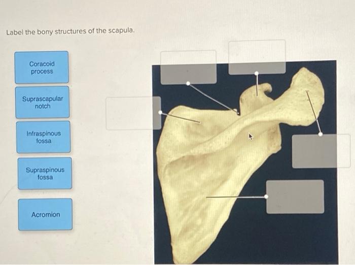
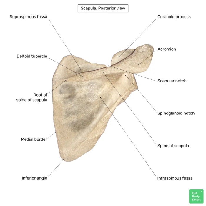
Label the bony structures of the scapula. – Label the bony structures of the scapula: a comprehensive overview that delves into the intricacies of this vital shoulder blade, exploring its anatomy, clinical significance, and imaging modalities. This exploration unravels the complexities of the scapula, providing a foundation for understanding its role in movement, stability, and injury.
The scapula, commonly known as the shoulder blade, is a flat, triangular bone situated in the posterior aspect of the shoulder. It serves as a critical attachment point for muscles involved in shoulder movement and stability, playing a pivotal role in various activities, ranging from simple daily tasks to complex athletic maneuvers.
Overview of the Scapula

The scapula, commonly known as the shoulder blade, is a flat, triangular bone located on the dorsal aspect of the thoracic cage. It serves as the primary attachment point for the upper limb and plays a crucial role in shoulder movement and stability.
The scapula’s complex anatomy and biomechanics make it clinically significant in various musculoskeletal conditions and injuries.
Bony Structures of the Scapula

The scapula comprises several distinct bony structures, each with its unique shape, size, and location:
Body
The body is the main, flattened portion of the scapula, providing attachment sites for various muscles. It is bounded by the superior, lateral, and medial borders, with the spine running along its posterior aspect.
Spine
The spine is a bony ridge that projects posteriorly from the body. It serves as an attachment site for the trapezius muscle and acts as a lever arm for scapular movement.
Acromion, Label the bony structures of the scapula.
The acromion is a flattened, hook-shaped process that extends laterally from the spine. It articulates with the clavicle to form the acromioclavicular joint.
Coracoid Process
The coracoid process is a beak-like projection located anterolateral to the scapular notch. It provides attachment for muscles involved in shoulder flexion and abduction.
Glenoid Cavity
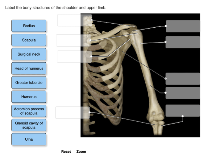
The glenoid cavity is a shallow, concave depression located on the lateral aspect of the scapula. It articulates with the head of the humerus to form the glenohumeral joint.
Clinical Relevance: Label The Bony Structures Of The Scapula.

The bony structures of the scapula play a significant role in movement, stability, and injury:
Movement
The scapula undergoes various movements, including elevation, depression, protraction, retraction, and rotation. These movements are essential for proper shoulder function and range of motion.
Stability
The scapula provides stability to the shoulder joint by anchoring the muscles that control its movement. The spine, acromion, and coracoid process serve as attachment points for muscles that stabilize the scapula and prevent excessive movement.
Injury
Injuries to the scapula can occur due to trauma, overuse, or underlying conditions. Common injuries include fractures, dislocations, and muscle tears. These injuries can affect shoulder function and cause pain, stiffness, and instability.
General Inquiries
What is the function of the scapula?
The scapula serves as an attachment point for muscles involved in shoulder movement and stability, facilitating a wide range of motions, including flexion, extension, abduction, adduction, and rotation.
What are the common injuries associated with the scapula?
Common injuries involving the scapula include fractures, dislocations, and muscle strains. Fractures can occur due to direct trauma or falls, while dislocations often result from high-energy injuries or sports-related accidents.
How is the scapula imaged?
The scapula can be visualized using various imaging modalities, including X-rays, CT scans, and MRI. X-rays provide a basic Artikel of the bone, while CT scans offer more detailed cross-sectional images. MRI is particularly useful for evaluating soft tissue structures surrounding the scapula.